NorrChemica™
Safranine O
Safranine O | CAS 477-73-6 | Dye Content: ≥85%
Couldn't load pickup availability
Technical Specifications
| CAS Number | 477-73-6 |
| EC / EINECS Number | 207-518-8 |
| MDL Number | MFCD00011759 |
| RTECS Number | SG1623000 |
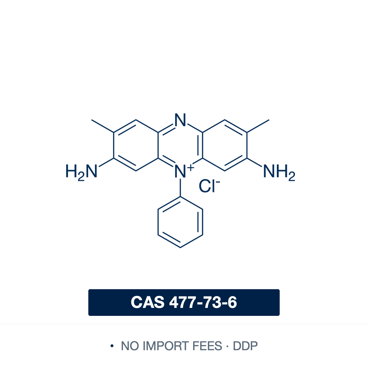
| SMILES | CC1=CC2=C(C=C1N)[N+](=C3C=C(C(=CC3=N2)C)N)C4=CC=CC=C4.[Cl-] |
| InChI | InChI=1S/C20H18N4.ClH/c1-12-8-17-19(10-15(12)21)24(14-6-4-3-5-7-14)20-11-16(22)13(2)9-18(20)23-17;/h3-11H,1-2H3,(H3,21,22);1H |
| InChIKey | OARRHUQTFTUEOS-UHFFFAOYSA-N |
| PubChem CID | 2723800 |
| Molecular Formula | C₂₀H₁₉ClN₄ |
| Molecular Weight | 350.85 g/mol |
| Solubility | ~50 mg/mL in water (20 °C); soluble in ethanol |
| Purity | Dye content: ≥85% |
| Physical Form | Dark-red crystalline powder |
| HS Code | 3204.13 |
| Country of Origin | Finland |
| Shelf Life | Retest period: 36 months from date of manufacture |
| Storage Conditions | Store in a cool, dry place in a tightly sealed container, protected from light. |
Product Description & Scientific Applications
Safranine O (Safranin O; C.I. 50240; Basic Red 2) is a cationic phenazinium dye used in histology, cytology, microbiology, and bioenergetics. As a basic dye it binds electrostatically to anionic cellular and extracellular components.
Microbiology counterstain. In the Gram stain, after crystal violet–iodine and decolourisation with ethanol, acetone, or ethanol/acetone, it re-stains decolourised Gram-negative cells pink to red while Gram-positive cells retain the crystal violet–iodine purple. In Schaeffer–Fulton endospore staining it counterstains the vegetative cells of Bacillus and Clostridium pink after malachite green is heat-driven into the endospore, giving the green-spore / red-cell contrast.
Cartilage and proteoglycan histology. Its cationic charge binds the anionic chondroitin and keratan sulfate chains of aggrecan, and orange-to-red intensity tracks sulfated-glycosaminoglycan content under controlled fixation, decalcification, and staining. Paired with Fast Green FCF, which counterstains bone and non-cartilage matrix, the Safranine-O / Fast Green protocol is used for OARSI and Mankin scoring of cartilage degradation and chondrocyte morphology in osteoarthritis — with subchondral-bone sclerosis read from the same counterstained sections — and as a chondrogenic-differentiation readout for mesenchymal stem cells. Glycosaminoglycan loss during decalcification attenuates intensity, so reference sections and controls accompany quantitation.
Plant histology. It stains lignified and suberised cell walls bright red, while Fast Green FCF, Alcian Blue, or Astra Blue stain cellulosic and non-lignified tissue green or blue — used to examine vascular differentiation, xylem and phloem, wood and bark anatomy, secondary cell-wall deposition, cuticle and periderm, and lignification in root, stem, and leaf.
Mitochondrial membrane potential. As a delocalised lipophilic cation it accumulates in energised mitochondria in a potential-dependent manner; stacking on anionic inner-membrane sites produces the absorbance shifts and fluorescence quenching that read out ΔΨm in isolated mitochondria and digitonin-permeabilised cells. It is recorded alongside oxygen consumption in combined high-resolution respirometry/fluorometry to probe respiratory-chain coupling and uncoupling.
Photosensitiser and redox mediator. It absorbs at 530–534 nm in 50% aqueous ethanol and is reversibly reduced to its colourless leuco form, leucosafranine; its two-electron midpoint potential is near −280 mV vs SHE at pH 7.0, though commercial Safranine O is an isomer mixture whose components span roughly −272 to −315 mV. It serves as an electron-transfer mediator in potentiometric titrations of low-potential biological redox couples, a colorimetric indicator in reduction-potential determinations, a visible-light sensitiser in dye-sensitised hydrogen photoproduction and electron-transfer studies, and a sensitiser/initiator in dye-sensitised photopolymerisation.
Other uses. Discrimination of connective-tissue from mucosal mast cells by sequential Alcian Blue / Safranine O — heparin-rich connective-tissue mast cells stain red, mucosal mast cells blue; model cationic dye in adsorption studies of dye removal by clay, hydrogel, polymeric, and nanocomposite adsorbents; model/target dye in TiO₂ and other semiconductor photocatalytic-degradation studies.
Shipping Destinations
- EU & UK: Priority delivery, 2–5 business days.
- United States (DDP): 3–7 business days, duties and taxes prepaid.
- EFTA Countries (DDP): 3–7 business days, duties and taxes prepaid.
- Worldwide: 7–14 business days, selected locations.
Safety Information
| GHS Pictograms |

|
| Signal Word | Warning |
| Hazard Class | None — not subject to transport regulations |
| Transport Category | Not classified as dangerous goods for transport (ADR/IATA/IMDG) |
| H-Statements | H315 - H319 |
| P-Statements | P280 - P302+P352 - P305+P351+P338 - P332+P313 - P337+P313 - P501 |
Documentation
| Safety Data Sheet | Download PDF |