NorrChemica™
Toluidine Blue O
Toluidine Blue O | CAS 92-31-9 | Dye Content ≥75%
Couldn't load pickup availability
Technical Specifications
| CAS Number | 92-31-9 |
| EC / EINECS Number | 202-146-2 |
| MDL Number | MFCD00011934 |
| RTECS Number | SP5670000 |
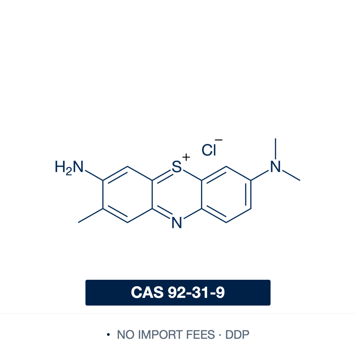
| SMILES | CC1=CC2=C(C=C1N)SC3=CC(=[N+](C)C)C=CC3=N2.[Cl-] |
| InChI | InChI=1S/C15H15N3S.ClH/c1-9-6-13-15(8-11(9)16)19-14-7-10(18(2)3)4-5-12(14)17-13;/h4-8,16H,1-3H3;1H |
| InChIKey | GEDVVYWLPUPJJZ-UHFFFAOYSA-N |
| PubChem CID | 7083 |
| Molecular Formula | C₁₅H₁₆ClN₃S |
| Molecular Weight | 305.83 g/mol |
| Melting Point | >200 °C (dec.) |
| Solubility | Soluble in water (~38 mg/mL), ethanol (~4 mg/mL), methanol; practically insoluble in xylen |
| Purity | Dye content ≥75% |
| Physical Form | Dark blue to dark green powder with bronze luster |
| HS Code | 3204.13 |
| Country of Origin | Finland |
| Shelf Life | Retest period: 36 months from date of manufacture |
| Storage Conditions | Store at room temperature in a tightly sealed container under inert atmosphere |
Product Description & Scientific Applications
Toluidine Blue O (tolonium chloride; C.I. Basic Blue 17; C.I. 52040) is a cationic thiazine/phenothiazinium dye used as a metachromatic stain in histology, microscopy, plant anatomy, and analytical staining. The monomer absorbs near 630 nm, giving orthochromatic blue staining of weakly anionic or nucleic-acid-rich components. On substrates of high anionic charge density — sulfated glycosaminoglycans, mast-cell granule proteoglycans, cartilage matrix — the polyanionic backbone screens cation-cation repulsion and the dye associates into close-packed face-to-face H-aggregates (dimers and higher), whose absorption shifts hypsochromically to 480–540 nm to give the characteristic red-purple metachromasia. The response strengthens with the substrate's anionic groups in the order carboxylate, then phosphate, then sulfate, and varies with dye concentration, ionic strength, and staining pH.
Mast-cell identification and metachromatic histology. Mast-cell granules are densely packed with sulfated proteoglycans, principally heparin, whose high charge density aggregates the dye on the granule to give red-purple metachromasia against a blue orthochromatic background of nuclei and cytoplasm. Acidic-pH protocols (typically pH 0.5–2.5 in citrate, acetate, or HCl buffers) sharpen the readout by leaving carboxylate-rich mucopolysaccharides unstained and reserving the dye for highly sulfated targets — underlying routine mast-cell counts in skin, gut, and respiratory-tract sections, mast-cell phenotyping in inflammation and connective-tissue research, and demonstration of strongly acidic mucopolysaccharides. After degranulation, reduced or redistributed granule metachromasia is itself a histochemical endpoint, since the local concentration and organisation of sulfated granule proteoglycans are altered.
Cartilage proteoglycan and extracellular-matrix assessment. In cartilage, the dye binds the anionic glycosaminoglycans and proteoglycans of the ground substance with high affinity for the sulfate residues of chondroitin and keratan sulfate: proteoglycan-rich territorial and interterritorial matrix stains metachromatic purple, while nuclei, cytoplasm, and surrounding matrix counterstain blue. This supports assessment of cartilage matrix organisation, proteoglycan distribution, chondrocyte differentiation in pellet and scaffold-seeded models, growth-plate morphology, and broader extracellular-matrix research. The same metachromatic principle drives quantitative dimethylmethylene blue and Toluidine Blue assays for sulfated glycosaminoglycan content in tissue digests, read photometrically from the absorbance shift between bound and free dye.
Rapid staining of semithin resin sections. It is the standard rapid stain for semithin (~0.5–1.0 μm) sections of epoxy- and acrylic-resin-embedded tissue in electron-microscopy workflows. Alkaline toluidine-blue/borate formulations rapidly stain heated semithin sections, the borate improving penetration into plastic-embedded material and giving strong contrast across nuclei, cytoplasm, extracellular matrix, basement membrane, and secretory or granular structures despite the low inherent contrast of resin sections under light microscopy. It is essential for orientation of blocks, region selection, and trim guidance before ultrathin sectioning, and for morphological evaluation of resin-embedded peripheral nerve, retina, kidney, and other small-block specimens where myelin, axon, and lamellar structures must be reviewed before TEM.
Plant anatomy and comparative cell-wall histochemistry. As a polychromatic stain, different cell-wall and cytoplasmic components give distinct colours in a single step: in mildly acidic to near-neutral buffers, lignified secondary walls of xylem vessels and sclerenchyma fibres appear blue-green to turquoise, pectin-rich primary walls and middle lamellae pink to magenta, cellulose and unlignified walls violet to blue, and nuclei and nucleoli deep blue. This differential response makes it the routine stain for surveys of xylem, phloem, sclerenchyma, vascular-bundle organisation, secondary growth, cell-wall differentiation, and suberised periderm, and for comparative morphology across vascular plants, bryophytes, and algae — often a one-step alternative to safranin/astra-blue or fast-green double staining.
Further applications.
- Nissl-substance and rough-endoplasmic-reticulum staining in neurohistology, where densely packed RNA-rich ribosomal arrays give the characteristic dark-blue Nissl pattern in neuronal cytoplasm.
- Photosensitiser in antimicrobial photodynamic-inactivation research against Gram-positive and Gram-negative bacteria, biofilms, and mixed communities under red-light irradiation, where excited-state Toluidine Blue O generates singlet oxygen and other reactive oxygen species that damage microbial membranes, proteins, and nucleic acids.
- Chromosome and chromatin staining in plant and animal cytogenetics, including squash and smear preparations.
- Acidic-glycocalyx and mucopolysaccharide visualisation in cell biology, including epithelial-mucin and cell-surface glycocalyx surveys.
Shipping Destinations
- EU & UK: Priority delivery, 2–5 business days.
- United States (DDP): 3–7 business days, duties and taxes prepaid.
- EFTA Countries (DDP): 3–7 business days, duties and taxes prepaid.
- Worldwide: 7–14 business days, selected locations.
Safety Information
| Hazard Class | None — not subject to transport regulations |
| Transport Category | Not classified as dangerous goods for transport (ADR/IATA/IMDG) |
Documentation
| Safety Data Sheet | Download PDF |
Share